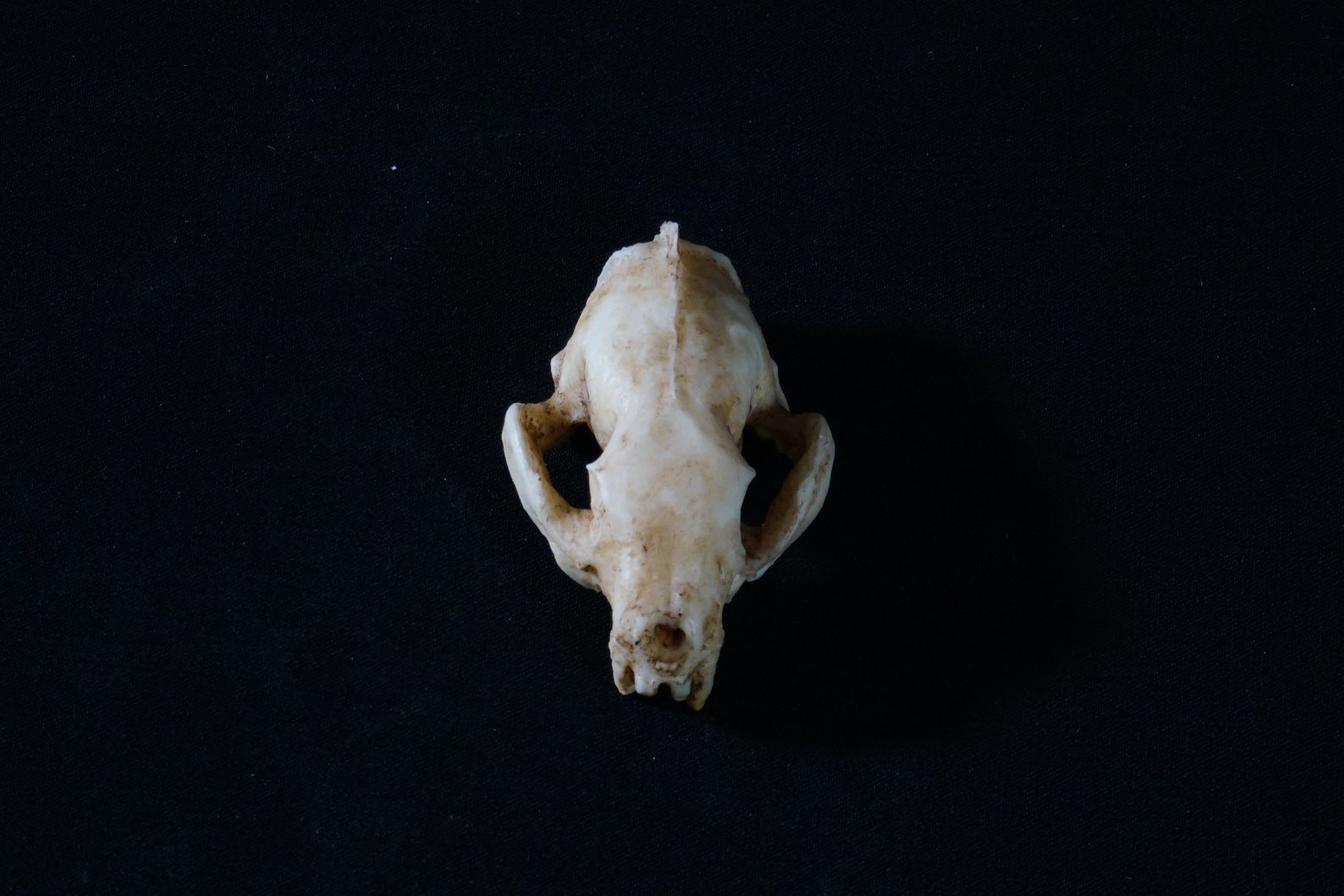
Ferret Skull Anatomy . Dorsal, lateral, ventral, caudal, cranial and midsagittal. The caudal crest (far caudal on skull) is a reliable and unambiguous skull. The skull was described macroscopically according to six standard views, i.e. Normal macroscopic and roentgenographic features of the skull of the ferret ( mustela putorius furo) were examined and described. Dorsal, lateral, ventral, caudal, cranial and midsagittal. Knowledge of the ferret skull’s intricate gross and radiographic anatomy is crucial for radiographic positioning,. Data were based on a sample of 100 (50 male and 50. For radiographic views of select pathologic conditions, anatomy, see chapter 35. This chapter discusses the anatomy of the domestic ferret (mustela putorius furo). The skull was described macroscopically according to six standard views, i.e. The following is a brief review of the clinically relevant anatomic and physiologic features of the ferret. The ferret's body shape and short limbs.
from www.etsy.com
Normal macroscopic and roentgenographic features of the skull of the ferret ( mustela putorius furo) were examined and described. This chapter discusses the anatomy of the domestic ferret (mustela putorius furo). Dorsal, lateral, ventral, caudal, cranial and midsagittal. Knowledge of the ferret skull’s intricate gross and radiographic anatomy is crucial for radiographic positioning,. The skull was described macroscopically according to six standard views, i.e. The caudal crest (far caudal on skull) is a reliable and unambiguous skull. The ferret's body shape and short limbs. The following is a brief review of the clinically relevant anatomic and physiologic features of the ferret. For radiographic views of select pathologic conditions, anatomy, see chapter 35. The skull was described macroscopically according to six standard views, i.e.
Ferret Resin Skull Reproduction Etsy
Ferret Skull Anatomy This chapter discusses the anatomy of the domestic ferret (mustela putorius furo). The following is a brief review of the clinically relevant anatomic and physiologic features of the ferret. The skull was described macroscopically according to six standard views, i.e. Dorsal, lateral, ventral, caudal, cranial and midsagittal. This chapter discusses the anatomy of the domestic ferret (mustela putorius furo). Data were based on a sample of 100 (50 male and 50. The skull was described macroscopically according to six standard views, i.e. Normal macroscopic and roentgenographic features of the skull of the ferret ( mustela putorius furo) were examined and described. Dorsal, lateral, ventral, caudal, cranial and midsagittal. For radiographic views of select pathologic conditions, anatomy, see chapter 35. Knowledge of the ferret skull’s intricate gross and radiographic anatomy is crucial for radiographic positioning,. The caudal crest (far caudal on skull) is a reliable and unambiguous skull. The ferret's body shape and short limbs.
From www.skullsunlimited.com
Replica BlackFooted Ferret Skull — Skulls Unlimited International, Inc. Ferret Skull Anatomy The following is a brief review of the clinically relevant anatomic and physiologic features of the ferret. Normal macroscopic and roentgenographic features of the skull of the ferret ( mustela putorius furo) were examined and described. The ferret's body shape and short limbs. Data were based on a sample of 100 (50 male and 50. Dorsal, lateral, ventral, caudal, cranial. Ferret Skull Anatomy.
From quizlet.com
Musteline skeletal anatomy of a ferret Diagram Quizlet Ferret Skull Anatomy Knowledge of the ferret skull’s intricate gross and radiographic anatomy is crucial for radiographic positioning,. The ferret's body shape and short limbs. The following is a brief review of the clinically relevant anatomic and physiologic features of the ferret. For radiographic views of select pathologic conditions, anatomy, see chapter 35. Data were based on a sample of 100 (50 male. Ferret Skull Anatomy.
From www.peepsburgh.com
Anatomy Of A Male Ferret PeepsBurgh Ferret Skull Anatomy The skull was described macroscopically according to six standard views, i.e. The ferret's body shape and short limbs. Knowledge of the ferret skull’s intricate gross and radiographic anatomy is crucial for radiographic positioning,. For radiographic views of select pathologic conditions, anatomy, see chapter 35. The skull was described macroscopically according to six standard views, i.e. Normal macroscopic and roentgenographic features. Ferret Skull Anatomy.
From www.peepsburgh.com
Ferret Internal Anatomy Labeled Ferret Skull Anatomy The skull was described macroscopically according to six standard views, i.e. The following is a brief review of the clinically relevant anatomic and physiologic features of the ferret. Dorsal, lateral, ventral, caudal, cranial and midsagittal. This chapter discusses the anatomy of the domestic ferret (mustela putorius furo). For radiographic views of select pathologic conditions, anatomy, see chapter 35. Data were. Ferret Skull Anatomy.
From www.flickr.com
comparison mink ferret skull John Rochester Flickr Ferret Skull Anatomy The skull was described macroscopically according to six standard views, i.e. Data were based on a sample of 100 (50 male and 50. The ferret's body shape and short limbs. For radiographic views of select pathologic conditions, anatomy, see chapter 35. Dorsal, lateral, ventral, caudal, cranial and midsagittal. Dorsal, lateral, ventral, caudal, cranial and midsagittal. The following is a brief. Ferret Skull Anatomy.
From www.deviantart.com
Ferret Skull by absol on DeviantArt Ferret Skull Anatomy For radiographic views of select pathologic conditions, anatomy, see chapter 35. This chapter discusses the anatomy of the domestic ferret (mustela putorius furo). The caudal crest (far caudal on skull) is a reliable and unambiguous skull. Dorsal, lateral, ventral, caudal, cranial and midsagittal. The ferret's body shape and short limbs. Dorsal, lateral, ventral, caudal, cranial and midsagittal. Knowledge of the. Ferret Skull Anatomy.
From www.vetexotic.theclinics.com
Ferret Oncology Veterinary Clinics Exotic Animal Practice Ferret Skull Anatomy Knowledge of the ferret skull’s intricate gross and radiographic anatomy is crucial for radiographic positioning,. Dorsal, lateral, ventral, caudal, cranial and midsagittal. The skull was described macroscopically according to six standard views, i.e. Data were based on a sample of 100 (50 male and 50. The following is a brief review of the clinically relevant anatomic and physiologic features of. Ferret Skull Anatomy.
From www.alamy.com
Skull Engraving Stock Photos & Skull Engraving Stock Images Page 3 Ferret Skull Anatomy The ferret's body shape and short limbs. Normal macroscopic and roentgenographic features of the skull of the ferret ( mustela putorius furo) were examined and described. The caudal crest (far caudal on skull) is a reliable and unambiguous skull. The skull was described macroscopically according to six standard views, i.e. The following is a brief review of the clinically relevant. Ferret Skull Anatomy.
From www.pinterest.com
Male FERRET Complete SKELETON Articulation Taxidermy Bone Oddity Ferret Skull Anatomy The skull was described macroscopically according to six standard views, i.e. Dorsal, lateral, ventral, caudal, cranial and midsagittal. This chapter discusses the anatomy of the domestic ferret (mustela putorius furo). The following is a brief review of the clinically relevant anatomic and physiologic features of the ferret. Data were based on a sample of 100 (50 male and 50. The. Ferret Skull Anatomy.
From www.pinterest.co.uk
Blackfooted Ferret Skull Animal Skeletons, Animal Skulls, Photo Ferret Skull Anatomy Normal macroscopic and roentgenographic features of the skull of the ferret ( mustela putorius furo) were examined and described. The caudal crest (far caudal on skull) is a reliable and unambiguous skull. The skull was described macroscopically according to six standard views, i.e. Knowledge of the ferret skull’s intricate gross and radiographic anatomy is crucial for radiographic positioning,. Dorsal, lateral,. Ferret Skull Anatomy.
From www.vetexotic.theclinics.com
Ferret Respiratory System Clinical Anatomy, Physiology, and Disease Ferret Skull Anatomy The skull was described macroscopically according to six standard views, i.e. The following is a brief review of the clinically relevant anatomic and physiologic features of the ferret. For radiographic views of select pathologic conditions, anatomy, see chapter 35. This chapter discusses the anatomy of the domestic ferret (mustela putorius furo). The caudal crest (far caudal on skull) is a. Ferret Skull Anatomy.
From www.pinterest.com
Ferret anatomy … Pinteres… Ferret Skull Anatomy Dorsal, lateral, ventral, caudal, cranial and midsagittal. The ferret's body shape and short limbs. The skull was described macroscopically according to six standard views, i.e. The following is a brief review of the clinically relevant anatomic and physiologic features of the ferret. For radiographic views of select pathologic conditions, anatomy, see chapter 35. Normal macroscopic and roentgenographic features of the. Ferret Skull Anatomy.
From pinterest.com
Black footed ferret skeleton structure 4H projects Pinterest Ferret Skull Anatomy Dorsal, lateral, ventral, caudal, cranial and midsagittal. For radiographic views of select pathologic conditions, anatomy, see chapter 35. The caudal crest (far caudal on skull) is a reliable and unambiguous skull. Knowledge of the ferret skull’s intricate gross and radiographic anatomy is crucial for radiographic positioning,. The following is a brief review of the clinically relevant anatomic and physiologic features. Ferret Skull Anatomy.
From www.etsy.com
Discount Domestic Ferret Skull Real Bone Animal by EvasFeathers Ferret Skull Anatomy The skull was described macroscopically according to six standard views, i.e. The following is a brief review of the clinically relevant anatomic and physiologic features of the ferret. The caudal crest (far caudal on skull) is a reliable and unambiguous skull. The skull was described macroscopically according to six standard views, i.e. Dorsal, lateral, ventral, caudal, cranial and midsagittal. The. Ferret Skull Anatomy.
From a-z-animals.com
Do Ferrets Have Spines? AZ Animals Ferret Skull Anatomy Dorsal, lateral, ventral, caudal, cranial and midsagittal. Dorsal, lateral, ventral, caudal, cranial and midsagittal. The skull was described macroscopically according to six standard views, i.e. Normal macroscopic and roentgenographic features of the skull of the ferret ( mustela putorius furo) were examined and described. The ferret's body shape and short limbs. The caudal crest (far caudal on skull) is a. Ferret Skull Anatomy.
From www.pinterest.com
This is great, so hard to find good diagrams of a ferret skeleton Ferret Skull Anatomy The skull was described macroscopically according to six standard views, i.e. The ferret's body shape and short limbs. The skull was described macroscopically according to six standard views, i.e. Knowledge of the ferret skull’s intricate gross and radiographic anatomy is crucial for radiographic positioning,. For radiographic views of select pathologic conditions, anatomy, see chapter 35. Data were based on a. Ferret Skull Anatomy.
From www.skullsunlimited.com
Replica BlackFooted Ferret Skull For Sale Skulls Unlimited Ferret Skull Anatomy The skull was described macroscopically according to six standard views, i.e. This chapter discusses the anatomy of the domestic ferret (mustela putorius furo). Normal macroscopic and roentgenographic features of the skull of the ferret ( mustela putorius furo) were examined and described. Knowledge of the ferret skull’s intricate gross and radiographic anatomy is crucial for radiographic positioning,. Dorsal, lateral, ventral,. Ferret Skull Anatomy.
From www.firstdensitymaterial.com
Load image into Gallery viewer, Ferret Skull (STL) Commercial License Ferret Skull Anatomy Dorsal, lateral, ventral, caudal, cranial and midsagittal. Dorsal, lateral, ventral, caudal, cranial and midsagittal. The skull was described macroscopically according to six standard views, i.e. Knowledge of the ferret skull’s intricate gross and radiographic anatomy is crucial for radiographic positioning,. This chapter discusses the anatomy of the domestic ferret (mustela putorius furo). The caudal crest (far caudal on skull) is. Ferret Skull Anatomy.